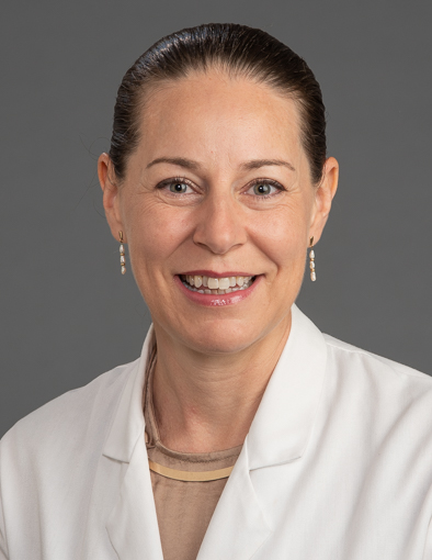
The Aneurysm and AVM Foundation is pleased to announce the recipients of the 2022 Cerebrovascular Research Grant Awards. We selected three researchers whose scientific projects showed the greatest potential to improve our understanding of cerebrovascular diseases.
 Research Study: Prevention Of Rbpj/Notch Mediated Brain Arteriovenous Malformation Via Blockade Of Apelin Or Osteopontin Signaling In Mice
Primary Investigator: Corinne M. Nielsen, Assistant Professor, Ohio University
Background: Dr. Nielsen’s team will use a genetic mouse model to study the mechanisms of mammalian brain arteriovenous malformation (AVM) disease formation and progression. To induce brain AVMs, they use a controllable gene deletion system to delete Rbpj, a gene that produces a protein that regulates expression of other “downstream” genes, in a type of molecular domino chain reaction. In the genetic mouse system they control the specific cells in which Rbpj is deleted, as well as the specific time at which Rbpj is deleted. Genetic deletion of Rbpj from endothelial cells leads to clinically defined features of brain AVM within two weeks. Based on the lab’s studies about how Rbpj-mutant brain AVMs develop, they identified molecules that may be targeted in attempts at preventing or treating hallmarks of brain AVMs.
Brain endothelial cells are isolated from one-week old control and mutant mice – from those cells, they identified genes that were up- or down- regulated, in mutants, as compared to controls. Among differentially expressed genes, two candidates showed increased gene expression in mutant brain endothelial cells. These molecules have been shown to regulate activation of small GTPases – molecules that help control cell shape, cell directionality, and cell migration, as part of a molecular “chain reaction” previously mentioned. These P7 expression studies demonstrated that the activity of a select small GTPase was likewise increased in mutant brain endothelium, at P7.
Because elevated expression and function of the molecules encoded by Apelin and Spp genes may act as mediators of the Rbpj-mutant brain AVM phenotype, Dr. Nielsen’s team hypothesizes that blocking cellular signaling through these molecules will prevent Rbpj-mediated brain AVMs. Their experimental plan is to induce their genetic manipulation, administer a reagent that blocks cellular signaling through each of the candidate molecules, and to assess features of brain AVMs, including vessel diameter, endothelial cell shape index, vessel permeability (leakage), and activation of small GTPases.
Research Objective: The goal of Dr. Nielsen’s proposal is the prevention of Rbpj/Notch mediated brain arteriovenous malformation via blockade of Apelin or Osteopontin signaling in mice..
Outcomes: Using the grant from the Aneurysm and AVM Foundation, Dr. Nielsen’s team hypothesizes that blocking cellular signaling at a pre-AVM time point will prevent the formation of brain AVMs in the Rbpj mutant mice. These prevention experiments will pave the way for future rescue experiments, which will determine whether blocking these molecules, after brain AVMs have already formed, will reverse features of brain AVMs.
Source: Corinne M. Nielsen, Assistant Professor, Ohio University. This research summary has been adapted and edited from Dr. Nielsen’s research proposal.
 Research Study: Piezo1 mechanoreceptor overexpression and dysregulation as a novel mechanism of intracranial aneurysm development
Primary Investigator: Stacey Q Wolfe, MD FAANS, Associate Professor, Residency Program Director, Wake Forest School of Medicine
Background: Intracranial aneurysms (IAs) are a life-threatening disease which still have no medical prevention or treatment. We still have a limited understanding of how they develop, though there does seem to be a link to high blood pressure and inflammation. Last year, The Nobel prize in Physiology was given to the scientist who discovered mechanoreceptors, special channels in the cell wall that sense pressure. In their preliminary studies of the actual aneurysm wall from 4 patients, Dr. Wolfe’s team found that these mechanoreceptors, called Piezo1 ion channels, were present in elevated numbers, much higher than in normal scalp arteries.
Additionally, they found that the pattern of organization in the aneurysm was lost, compared to normal arteries. This change in number and pattern of Piezo1 mechanoreceptors is significant because it leads to altered response to pressure, which may lead to development or rupture of IA. Furthermore, Piezo1 receptors are found to activate immune cells like macrophages to a pro-inflammatory state, and inflammation is known to cause aneurysm wall degeneration.
Dr. Wolfe’s team will examine the aneurysm wall collected during routine aneurysm clipping surgery of 25 patients to evaluate for overexpression and dysregulation of this receptor, and assess its location and how it influences the artery wall and associated inflammatory changes. They will also look for a common mutation of this that is found in certain ethnicities to better understand its function and differences in various populations.
Research Objective: The goal of Dr. Wolfe’s proposal is to demonstrate expression of the Piezo1 mechanoreceptor ion channel in human intracranial aneurysm tissue specimens and measure the presence and effect of E756 deletion on the histopathology of human intracranial aneurysms as a biologic variable.
Outcomes: With the grant from the Aneurysm and AVM Foundation, Dr. Wolfe’s team hope that their study evaluating the Piezo1 mechanism, the first study of its kind, can lead to a medical treatment to prevent development and rupture of aneurysms by using medications that regulate these mechanoreceptors.
Source: Stacey Q Wolfe, MD FAANS, Associate Professor, Residency Program Director, Wake Forest School of Medicine. This research summary has been adapted and edited from Dr. Wolfe’s research proposal.
 Research Study: Detection of activating somatic mutations in vascular malformations using minimally-invasive techniques
Primary Investigator: Ann Mansur MD, PhD Candidate, Research Trainee (Neurosurgery Resident and PhD Candidate), Research Financial Services, University Health Network
Background: Vascular malformations (VMs) are abnormal vessels in the body that include arteriovenous malformations (AVMs), venous malformations (VeMs) and lymphatic malformations (LMs). These abnormal vessels are prone to bleeding and can grow significantly; they cause hemorrhage, seizures, neurological deficits, deformity and pain. Despite the availability of traditional therapies including surgery, radiation and endovascular procedures, many of these VMs are insufficiently treated, causing tremendous morbidity and mortality to patients.
In the past decade, focused research has been conducted on understanding the molecular and genetic bases of these VMs. Scientists have found that these lesions are caused by specific mutations that occur in a non-hereditary way. There are now novel oral medications being researched to target these mutations in efforts to gain a new treatment strategy to battle these complex diseases. The identification of these mutations requires a tissue biopsy, which is not only invasive, but also requires a specialized neurosurgeon, and can cause inadvertent complications including bleeding, infection and wound issues.
A “liquid biopsy” involves (1) sampling blood or cyst fluid in a minimally invasive manner and (2) using novel molecular techniques to extract the mutations from the fluid. This technique has been increasingly adopted in cancer populations with good success and significantly less risk. A few isolated case reports in the past two years have shown that while sampling peripheral blood in patients with VMs doesn’t sufficiently detect these mutations, sampling blood/cyst fluid closer to the lesion or sampling the wall of the lesion at the time of an endovascular procedure can detect various clinically relevant mutations with minimal to no additional risk.
Research Objective: The goal of Dr. Mansur’s proposal is to validate the novel technique of cell-free DNA (ctDNA) next-generation sequencing (NGS) liquid biopsies in detecting targeted mutations in vascular malformations.
Outcomes: Using this grant from the Aneurysm and AVM Foundation, Dr. Mansur’s team hope to validate these new minimally invasive diagnostic approaches in hopes to establish a reliable and feasible way to determine a patient’s VM mutations, and perhaps their candidacy for targeted oral medications for these specific mutations.
Source: Ann Mansur MD, PhD Candidate, Research Trainee (Neurosurgery Resident and PhD Candidate). This research summary has been adapted and edited from Dr. Mansur’s research proposal.
