Grant Recipients

The Aneurysm and AVM Foundation is pleased to announce the recipients of the 2025 Cerebrovascular Research Grant Awards. We selected four researchers whose scientific projects showed the greatest potential to improve our understanding of cerebrovascular diseases.
Research Study: Promoting brain hematoma clearance via immunomodulation of infiltrating macrophages.

Primary Investigator: Sahily Reyes-Esteves, MD, PhD, Instructor/Incoming Assistant Professor (CE Track), University of Pennsylvania

Background: Intracranial hemorrhage (ICH) is a deadly form of stroke caused by bleeding in the brain with minimal therapeutic strategies. Among ICH causes, ruptured vascular malformations disproportionately affect younger patients and thus, lead to a significant loss of productive life- years. The recent success of surgical hematoma evacuation trials in ICH suggests that strategies that decreased hematoma burden could be beneficial for patients with ICH, but many are not surgical candidates.

Nanoparticles have been clinically available for decades, including the successful mRNA COVID vaccine packaged in lipid nanoparticles (LNPs). Given LNP’s potential for delivering a variety of cargo to specific cell types, bioengineering of CNS-targeted LNPs could provide powerful therapy for ICH of multiple etiologies, including ruptured vascular malformations. Using a mouse model of ICH, Dr. Reyes-Esteves has developed a vehicle for drugs using tiny drug carriers called lipid nanoparticles (LNPs) to deliver treatment molecules to these macrophages.

Using the ICH model in mice, it was noted that LNPs conjugated to antibodies targeting adhesion molecules (such as the vascular cell adhesion molecule, VCAM) resulted in improved brain drug delivery compared to passive delivery, even at times of high vascular leakage. Based on this, the team built a platform technology for drug delivery to the brain during ICH, which achieved 5x higher brain delivery compared to passive delivery. This proves that reduction in hematoma size is a feasible therapeutic target after ICH that leads to behavioral improvements, and sets the stage for the central hypothesis that LNP-mediated immunomodulation of brain- infiltrating macrophages after ICH can improve hematoma phagocytosis and outcomes in experimental ICH. 

Research Objective: The goal of Dr. Reyes-Esteves’ proposal is to develop a new treatment that will use molecular tools to encourage macrophages to remove blood more effectively from the brain while reducing harmful inflammation.

Outcomes: With this grant from the Aneurysm and AVM Foundation, Dr. Reyes-Esteves hopes this research could lead to a non-invasive therapy that helps more ICH patients recover with fewer complications.

Source: Dr. Sahily Reyes-Esteves, MD, PhD. This research summary has been adapted and edited from Dr. Reyes-Esteves’ research proposal.

Research Study: Trial of Small Molecule Inhibitors in a Novel, Localized, Inducible, Sporadic Brain Arteriovenous Malformation Mouse Model.

Primary Investigator: Dr. Rashad Jabarkheel, Department of Neurosurgery, Perelman School of Medicine, University of Pennsylvania

Background: As members of the TAAF community well know, the diagnosis of a brain AVM is devastating. Brain AVMs are complex vascular lesions where there is an abnormal tangle of blood vessels such that high flow arterial blood is directly shunted into veins, bypassing a normal capillary bed. Brain AVMs are high risk lesions because the abnormal blood flow within them makes them prone to rupture and consequently cause massive brain bleeds. Treatment of brain AVMs is challenging. For at least a third of brain AVMs, which are high grade, curative microsurgery is not an option, and any currently available treatment option is associated with poor outcomes. Unfortunately, no medications exist to decrease the size of brain AVMs and make them go away.

The goal of Dr. Jabarkheel’s project is to develop an effective medical treatment option for brain AVMs. It has recently been found that the majority of brain AVMs are due to mutations in a specific signaling pathway in the body called the KRAS pathway. Interestingly, the KRAS pathway is also involved in nearly half of all human cancers, and just in the past couple of years new KRAS pathway inhibitors have been developed for this purpose. This proposal wishes to test whether these KRAS pathway inhibitors can not only treat cancer, but also cause brain AVMs to regress.

To do so, Dr. Jabarkheel has developed a mouse model of brain AVMs that allows the team to monitor AVM size continuously. The mice will be injected with AAV-Dre through a cranial window at 6-8 weeks of age. The mice will be monitored for the development of AVMs by visible light imaging at post-operative day and fluorescein angiography for 4 weeks. The primary endpoint of the study is measured frequency of AVM regression.

Research Objective: The goal of Dr. Jabarkheel’s proposal is to trial whether KRAS pathway inhibitors can cause brain AVMs to regress in mice.

Outcomes: Using this grant from the Aneurysm and AVM Foundation, Dr. Jabarkheel hopes the research will set the stage for clinical trials of KRAS pathway inhibitors for treatment of human brain AVMs.

Source: Dr. Rashad Jabarkheel, Department of Neurosurgery. This research summary has been adapted and edited from Dr. Jabarkheel’s research proposal.

Research Study: Patient-Specific CFD Modeling of Intracranial Aneurysms: Distinguishing Hemodynamics of Aneurysms with and without Blebs with Inlet Boundary Conditions from 4D flow MRI.

Primary Investigator: Isaac Josh Abecassis, Assistant Professor, University of Louisville School of Medicine

Background: Intracranial aneurysms (IAs) can be devastating if they rupture, with almost half the patients dying and the remainder with significant disability. Sometimes IAs develop blebs or daughter sacs, which can indicate a spot of weakness in the aneurysm wall. Blebs can also develop on other cerebrovascular pathologies, like AVM draining veins. While computational fluid dynamics (CFD) can estimate important flow-related features that are associated with rupture, at least to date, most of these models do not factor in patient-specific biological conditions (i.e. how stiff the blood vessel wall is, how viscous the blood is, and the patient’s blood pressure). We aim to determine the difference in flow conditions between aneurysms with blebs from those without blebs.

4D-Flow magnetic resonance imaging (4DF-MRI) is a patient-specific, noninvasive MRI technique that collects detailed blood flow information from patients directly while inside an MRI scanner. This information can be used to inform more accurate CFD models; however, it has substantially lower resolutions. We propose to develop high resolution CFD models which utilize 4DF-MRI in patients with IAs, with an emphasis on the presence of blebs, resulting in highly accurate flow-related parameters (like Wall Shear stress (WSS) and Oscillatory Shear Index (OSI)). Understanding flow conditions within IAs and near blebs will help clinicians triage “higher risk” versus “lower risk” aneurysms.

Ultimately, the results of this investigation will help to develop a real-time “CFD-like” analysis on aneurysms, blebs associated with aneurysms, and AVM draining veins with and without venous varices to determine the risk of bleeding. Future studies will also focus on specific aneurysm locations that are more prone to rupture (i.e. Anterior communicating artery) to determine if these flow- related parameters are different based on location.

Research Objective: The goal of Dr. Abecassis’ proposal is to develop patient-specific CFD models based on 4DF-MRI in patients with IAs with and without blebs, and derive key parameters that have shown prognostic value in predicting aneurysm rupture. 

Outcomes: With this grant from the Aneurysm and AVM Foundation, Dr. Abecassis’ hopes this can prompt more studies in the future to determine if these flow- related parameters are different based on location.

Source: Dr. Isaac Josh Abecassis, Assistant Professor. This research summary has been adapted and edited from Dr. Abecassis’ research proposal.


Research Study: Precision Gene Correction for Sporadic KRAS-Driven Brain Arteriovenous Malformations.

Primary Investigator: Pazhanichamy Kalailingam, PhD, Harvard Medical School

Background: Brain arteriovenous malformations (bAVMs) are high-flow anomalous connections between cerebral arteries and veins that bypass the intervening capillary network, leading to a high risk of intracranial hemorrhage, often causing death or lifelong disability.Sporadic brain AVMs (bAVM), affecting approximately 15 per 100,000 people per year in the US2,, are often caused by somatic mutations, particularly in the KRAS and BRAF genes in > 60% of cases. Treatment options include open neurosurgical resection, endovascular embolization, and stereotactic radiosurgery (SRS) but are often ineffective for large and eloquent lesions.

Hemorrhage of bAVMs can be lethal, with a ~4%15 annual rupture rate (compounded over one’s lifetime, as most bAVM are diagnosed in childhood), and often go undetected until symptomatic. Moreover, residual deficits after hemorrhage include epilepsy, complex headache, cognitive and pain syndromes. Without effective treatments, bAVMs remain “ticking time bombs,” underscoring the urgent need for non-surgical, molecular-targeted therapies.

Somatic gain-of-function mutations in KRAS and BRAF have been identified in 60–81% of sporadic bAVM, are present at low allele frequencies (0.5–4%), and are primarily enriched in endothelial cell (EC) populations. These mutations disrupt EC function, cause abnormal EC size and migration, together driving bAVM formation and predisposition to hemorrhage. Among these mutations, the KRASG12D mutation is the most prevalent (32-52.4% of cases; ~30,000 patients in the USA), making it an ideal target for therapeutic intervention. While small molecule therapies targeting the KRAS pathway, such as those developed for cancer, show high toxicity due to lack of selectivity for mutant KRAS, recent allosteric inhibitors for KRASG12C have shown promise in reducing AVM size (extracranial) in limited cases. These studies demonstrate the feasibility of inhibiting KRAS activity as a viable strategy for bAVM treatment in humans.

Research Objective: The goal of Dr. Kalailingam’s proposal is to optimize CRISPR base editing to correct the KRASG12D mutation in human and mouse endothelial cells, restoring normal function and reducing in vivo AVM formation.

Outcomes: Using this grant from the Aneurysm and AVM Foundation, Dr. Kalailingam hopes this research can lay the foundation for future translational efforts to develop targeted gene therapies for AVM patients, addressing an urgent, unmet clinical need.

Source: Dr. Pazhanichamy Kalailingam Senior Scientist. This research summary has been adapted and edited from Dr. Kalailingam’s research proposal.

The Aneurysm and AVM Foundation is pleased to announce the recipients of the 2024 Cerebrovascular Research Grant Awards. We selected four researchers whose scientific projects showed the greatest potential to improve our understanding of cerebrovascular diseases.

Research Study: A novel tyrosine kinase inhibitor coated flow diverting stent as a drug delivery system for intracranial aneurysms

Primary Investigator: Malia McAvoy, MD MS, Resident Physician, University of Washington

Background: Currently, doctors use less invasive techniques to treat brain aneurysms. They do this by reducing blood flow to the weakened area to prevent it from getting larger. This research will develop a new approach to improve aneurysm treatment. The objective is to develop a special device that slows down blood flow and also uses particular drugs to target the main genetic factors that lead to aneurysms. This device, called a stent, will carry medicine on its surface to address the genetic and biological causes of aneurysm in the brain where they occur.

This approach is newly possible because of an innovative coating technique Dr. McAvoy has developed that will allow us to place therapeutic drugs on the surface of stents and have the stent release drugs over a long period of time. With this new device, patients can avoid the negative side effects that often occur with various drug treatments, and at the same time receive treatments that target the root cause of the aneurysm.

In this research project, the first step is to coat stents with drugs and confirm that the drug coating is stable on the stent surface. The second step is to measure how quickly or slowly the drug releases from the surface of the stent. This will be done using 3D-printed models of actual patient aneurysms and simulating blood flow.

Research Objective: The goal of Dr. McAvoy’s proposal is to test if localized TKI drug delivery from the surface of existing endovascular devices currently used to treat IAs could be used to treat fusiform IAs as an innovative treatment solution.

Outcomes: With this grant from the Aneurysm and AVM Foundation, Dr. McAvoy’s hopes this project can help to develop a new treatment approach that will permit personalized and targeted drug delivery for the treatment of brain aneurysms. 

Source: Malia McAvoy, MD MS, Resident Physician. This research summary has been adapted and edited from Dr. McAvoy’s research proposal.


Research Study: Targeting cell adhesion molecules for treating brain arteriovenous malformation
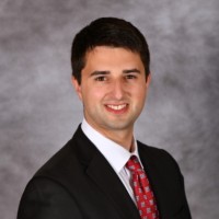
Primary Investigator: Eunsu Park, Assistant Professor, The University of Texas Health Science Center at Houston

Background: Brain arteriovenous malformations (bAVM) often cause intracerebral hemorrhages (ICH), which are associated with a 40-52% rate of mortality and morbidity. Surgical removal of bAVMs is an applicable treatment option; however, this may lead to significant damage to normal brain tissues in the case of deep brain AVMs. Dr. Park’s goal is to develop pharmacological treatment options for inhibiting bAVM-caused ICH.

Clinical studies report that up to 63% of human bAVM patients harbor KRAS mutations in the endothelial cells. KRAS is a family of small guanosine triphosphatases (GTPases) that is a binary molecular switch to  turn on/off genes related to cell cycle, differentiation, or survival. The KRAS mutation is detected explicitly in brain vascular ECs. Based on the evidence, Dr. Park has established a novel bAVM mouse model by inducing KRASG12V mutation in brain ECs using brain EC specific AAV capsid. The KRASbAVM mice model recapitulates human bAVM pathology, including tangled vessels, spontaneous ICH, and neurological deficits. However, the mechanisms by which KRAS mutations cause ICH remain unclear.

This leads to their central hypothesis that inhibition of CAMs reduces M/M activation/infiltration, preventing ICH in KRASbAVM mice. This will be tested through long-term & simultaneous inhibition of CAMs to test if the long-term VCAM1 or simultaneous CAM inhibition reduces M/M activation/infiltration and ICH. Dr. Park's team will also determine the fine stereotaxic coordinate of bAVM nidus using MRA and mouse brain atlas by injecting a cocktail of CAM MAbs into the bAVM nidus in KRASbAVM mice, testing the ICH using MRI/A and histology.

Research Objective: The goal of Dr. Park’s proposal is to uncover a causative role of CAMs in M/M activation/migration, causing bAVM rupture/ICH and will provide evidence for a pharmacological strategy for inhibiting bAVM-caused ICH.

Outcomes: Using this grant from the Aneurysm and AVM Foundation, the completion of Dr. Park’s preclinical study hopes to be useful in the development of an inflammation-modulation strategy to stabilize bAVM in patients.

Source: Eunsu Park, Assistant Professor, The University of Texas Health Science Center at Houston. This research summary has been adapted and edited from Dr. Park’s research proposal.


Research Study: A Prospective Study Examining Mobility and Sleep after Aneurysmal Subarachnoid Hemorrhage using a Wearable Fitness Tracker
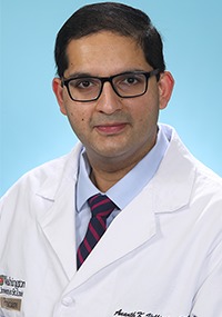
Primary Investigator: Matthew K. McIntyre, MD, Neurosurgery Resident, Oregon Health and Science University, Portland, OR

Background: Aneurysmal subarachnoid hemorrhage (aSAH) carries high long-term morbidity. The vast majority of aSAH research focuses on the inpatient management of this disease with the frequent classification of a ‘good outcome’ as a modified Rankin score (mRS) of ≤2. However, even among this group of functionally-independent aSAH patients, nearly two thirds report  dissatisfaction and participation restrictions with their daily activities especially with housekeeping, chores, and physical exercise at 6 months post ictus. At 1 year, 33% of aSAH patients report ongoing issues with fatigue, cognitive problems, and emotional problems that are associated with poorer mobility and learning.

Sleep dysfunction occurs in up to 37% of aSAH patients often affecting patients beyond one year post-ictus and is associated with poorer quality of life.This dysfunction likely has profound implications on return to work and functional  independence. It remains unclear what the burden of poor sleep is on a day-to-day basis for aSAH patients and what the implications of this deficit is on their functional outcomes after discharge. There is a growing body of evidence that high quality/quantity sleep is associated with improved learning, attention, physical activity, and recovery from brain injury, however this knowledge has not yet been leveraged for improving aSAH outcomes.  

Wearable technology, including commercially available fitness trackers such as those produced by Garmin, Fitbit, and Apple, offer unprecedented insight into a patient’s functional status. To date, wearable technology has been shown to identify early post-operative complications, increase physical activity, improve health-related cancer outcomes, and, in the spine literature, to improve post-operative mobility. At this time, we have little understanding of the ‘expected’ time to return to normal sleep patterns after SAH and others have gone on to suggest that there may be distinct subgroups of aSAH patients in terms of recovery patterns. Wearable devices will likely play an ever-growing role in the comprehensive evaluation of patient-centered health outcomes. The goal of Dr. McIntyre’s project is to leverage the emerging data behind sleep and mobility science to improve the functional outcomes of their aSAH patients.

Research Objective: The goal of Dr. McIntyre’s proposal is to establish the expected time to sleep and mobility recovery, develop a granular understanding of  the influence of in- and out of- hospital interventions on sleep and functional outcomes among aSAH patients, and to compare objective measures of mobility and sleep via a fitness watch with patient reported outcomes. 

Outcomes: With this grant from the Aneurysm and AVM Foundation, Dr. McIntyre hopes his research can help patients set realistic goals and to identify patients who are failing to meet these goals in order to allocate specific interventions such as sleep coaching and/or intensive physical therapy programs in order to improve their functional outcome after aSAH.  

Source: Matthew K. McIntyre, MD, Neurosurgery Resident. This research summary has been adapted and edited from Dr. McIntyre’s research proposal.


Research Study: Role of IL-17 in Neurovascular Dysfunction after Subarachnoid Hemorrhage

Primary Investigator: Ananth K. Vellimana, Assistant Professor, Department of Neurological Surgery, Washington University in St. Louis

Background: Rupture of a brain aneurysm primarily leads to bleeding at the base of the brain and results in a condition called subarachnoid hemorrhage (SAH). Patients who experience SAH have a devastating outcome with  around 45% death within 30 days after the bleed and significant disability in nearly 50% of survivors. Two forms of secondary brain injury which are initiated by the aneurysm bleed, called Early Brain Injury (EBI) and Delayed Cerebral Ischemia (DCI) account for the majority of poor outcomes after SAH. While EBI occurs in the first few days after SAH, DCI typically occurs between 4-10 days after the initial bleed.

Recent studies have identified a variety of factors that lead to the development of EBI and DCI after SAH. Among these factors, inflammation in the blood, cerebrospinal fluid, and brain tissue plays a key role. A cytokine called IL-17 secreted by various cells of the immune system is a key contributor to the post SAH inflammatory response and has deleterious effects.

In this study, Dr. Vellimana will utilize a well-established mouse experimental model of SAH to understand whether inhibition of IL-17 using medications and genetically (via mice that lack IL-17) provides protection against EBI and DCI. If successful, additional studies will be performed in other animal models of SAH, with the ultimate goal of translating IL-17 directed treatment to patients with SAH. 

Research Objective: The goal of Dr. Vellimana’s proposal is to test the hypothesis that increased IL-17 production after SAH contributes to the development of EBI, DCI and long-term neurobehavioral dysfunction.

Outcomes: Using this grant from the Aneurysm and AVM Foundation, Dr. Vellimana hopes the research will be able to translate from experimental models to patients, as several IL-17 inhibitors are already FDA approved for conditions such as psoriatic arthritis and ankylosing spondylitis.

Source: Ananth K. Vellimana, Assistant Professor, Department of Neurological Surgery. This research summary has been adapted and edited from Dr. Vellimana’s research proposal.

The Aneurysm and AVM Foundation is pleased to announce the recipients of the 2023 Cerebrovascular Research Grant Awards. We selected three researchers whose scientific projects showed the greatest potential to improve our understanding of cerebrovascular diseases.

Research Study: Elucidating the genetic basis of Vein of Galen malformation

Primary Investigator: Andrew T. Hale, MD, PhD, Neurosurgery Resident, University of Alabama at Birmingham, Department of Neurosurgery

Background: Vein of Galen malformations (VOGMs) are the most common and severe neonatal blood vessel anomaly of the human  cerebral vasculature. VOGM are the result of abnormal connections between arteries, veins, and capillary vessels during neonatal development. Even with neurosurgical intervention, VOGM can cause systemic problems such as cardiac failure due to the heart’s inability to accommodate the largest amount of blood flow into the venous system. The VOGM may also cause increased intracranial pressure, leading to brain hemorrhage or impaired draining of cerebrospinal fluid resulting in hydrocephalus. These problems may be life-threatening.

Despite all of this, the cause of VOGM is largely unknown, but is widely hypothesized to be caused by genetic abnormalities. To  understand the genetic and molecular basis of VOGM, Dr. Hale proposes the creation of the VOGM Genetics Research Consortium (VOGM-GRC), an international multi-institutional network of the world’s leading pediatric neurosurgeons, endovascular specialists, vascular neurologists, and geneticists/molecular biologists. The team will aggregate genetic data from both germline (i.e., cheek swab DNA) as well as endoluminal biopsy of the VOGM lesion directly.

Understanding the genetic basis of VOGM may broadly inform our understanding of cerebrovascular development and disease pathogenesis; improve clinical prognostication and decision-making; and provide a mechanistic foundation for the rationale use of repurposed drugs and/or design of targeted therapeutics relevant for VOGM and potentially other cerebrovascular lesions.

Research Objective: The goal of Dr. Hale’s proposal is to identify causative genetic and molecular factors governing VOGM development and maintenance, with the goal of repurposing or developing novel pharmacologic therapies to improve VOGM treatment. 

Outcomes: With this grant from the Aneurysm and AVM Foundation, Dr. Hale’s team hopes their study can serve as an archetype for applying endoluminal biopsy techniques to pediatric cerebrovascular disease more broadly. 

Source: Andrew T. Hale, MD, PhD, Neurosurgery Resident, University of Alabama at Birmingham. This research summary has been adapted and edited from Dr. Hale’s research proposal.


Research Study: Enhancing glymphatic-lymphatic clearance as a novel strategy to treat SAH
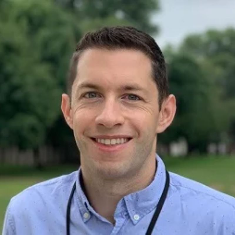
Primary Investigator: Humberto Mestre, Resident Physician, Department of Neurology, University of Pennsylvania

Background: Sadly, many aneurysms and arteriovenous malformations (AVM) are diagnosed too late, and they are often discovered after they have ruptured or bled causing a condition known as subarachnoid hemorrhage (SAH). While advances are being made in the earlier diagnosis of these conditions, SAH still remains one of the most common complications of aneurysms and AVM. In SAH, blood traveling in the cerebral blood vessels exits and accumulates around the brain in a space occupied by  the cerebrospinal fluid (CSF). Normally, CSF has a composition similar to tears (salty and translucent) and is produced by a specialized tissue in the brain called the choroid plexus. It possesses the perfect combination of water, minerals, and proteins for our brain cells to function properly and this is the fluid typically collected during spinal taps.

One recent discovery suggests that CSF continuously drains into the lymphatic system embedded into one of the linings of the brain called the dura, and this drainage acts as a waste disposal system, clearing out waste that accumulates during the day. After SAH, blood enters the CSF space and wreaks havoc, causing inflammation and adding to the damage caused by the bleed. The breakdown of blood causes blood vessels to clamp down, limiting the amount of blood flowing to the brain. It irritates brain cells causing them to be hyperactive leading to seizures, and it also clogs the drainage routes of CSF causing an abnormal accumulation of fluid known as hydrocephalus.

Current treatments after SAH are primarily focused towards preventing new bleeds and treating the complications, but none are targeted at removing blood from the CSF. But what if we could apply a painless, non-invasive ointment on the neck of patients after SAH to stimulate the removal of blood from the skull? Dr. Mestre’s proposal is based on a naturally occurring compound (prostaglandin F2α) that causes increased lymphatic drainage of CSF and could potentially harness our bodies’ natural abilities to speed up the removal of blood. Their hope is that this treatment could improve recovery and reduce the odds of developing complications after SAH.

Research Objective: The goal of Dr. Mestre’s proposal is to test a novel application of an FDA-approved prostanoid, prostaglandin F2α (PGF2α), in SAH which has been shown to augment transport along cervical lymphatic vessels and enhance CSF drainage in an age-independent manner. 

Outcomes: Using this grant from the Aneurysm and AVM Foundation, Dr. Mestre’s team hopes that their treatment strategy could not only improve the recovery of patients after SAH, but that it could also be expanded to treat other diseases such as stroke, traumatic brain injury, or even diseases like Alzheimer’s and Parkinson’s disease.  

Source: Humberto Mestre, Resident Physician, Department of Neurology, University of Pennsylvania. This research summary has been adapted and edited from Dr. Mestre’s research proposal.


Research Study: Overcoming barriers in Therapeutics, Imaging and Biomarkers for brain AVMs

Primary Investigator: Gael Genet, Research Assistant Professor, University of Virginia, School of Medicine, Cell Biology Dept

Background: Blood vessels are highly organized networks of arteries, capillaries, and veins. In patients with mutations that disrupt the mechanisms that regulate blood vessel formation – as in the genetic disease Hereditary Hemorrhagic Telangiectasia (HHT) – this can lead to the formation of abnormal, disorganized blood vessels (termed vascular malformations) that are prone to sudden and serious bleeding, particularly in the brain.

HHT affects 1 in 5,000 live births. Because it can vary in its clinical presentation, HHT can be difficult to diagnose. Symptoms often begin in childhood and progress in severity, but patients typically do not receive a definitive HHT diagnosis until >40 years of age. Drugs currently being used to treat that disease are helpful to many individuals but do not work for all patients and cause a lot of side effects. Therefore, it is needed to find new drug treatments to treat people afflicted with this illness.

Dr. Genet’s preliminary results showed that proper control of cell division (or cell cycle) is important for normal vessel formation. Their team found that uncontrolled cell division leads to blood vessel malformations. They will use new mouse models to study the modifications of the cell cycle in blood vessels in normal and disease conditions. They will also test the effects of drugs that are known to control cell cycle to prevent or cure vascular malformations, and develop new imaging techniques to visualize those malformations in the brain.

Research Objective: The goal of Dr. Genet’s proposal is to investigate dysregulation of endothelial cell cycle and identity in bAVMs, and validate the therapeutic potential of cell cycle modulators to treat vascular malformations. 

Outcomes: Using this grant from the Aneurysm and AVM Foundation, Dr. Genet’s project will provide new hope for the treatment of HHT patients, whereas current treatments are unsatisfactory and involve invasive surgery or radiation, with considerable risks and undesirable side-effects.

Source: Gael Genet, Research Assistant Professor, University of Virginia, School of Medicine, Cell Biology Dept. This research summary has been adapted and edited from Dr. Genet’s research proposal.

The Aneurysm and AVM Foundation is pleased to announce the recipients of the 2022 Cerebrovascular Research Grant Awards. We selected three researchers whose scientific projects showed the greatest potential to improve our understanding of cerebrovascular diseases.

Research Study: Prevention Of Rbpj/Notch Mediated Brain Arteriovenous Malformation Via Blockade Of Apelin Or Osteopontin Signaling In Mice
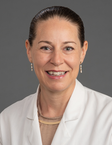
Primary Investigator: Corinne M. Nielsen, Assistant Professor, Ohio University

Background: Dr. Nielsen’s team will use a genetic mouse model to study the mechanisms of mammalian brain arteriovenous malformation (AVM) disease formation and progression. To induce brain AVMs, they use a controllable gene deletion system to delete Rbpj, a gene that produces a protein that regulates expression of other “downstream” genes, in a type of molecular domino chain reaction. In the genetic mouse system they control the specific cells in which Rbpj is deleted, as well as the specific time at which Rbpj is deleted. Genetic deletion of Rbpj from endothelial cells leads to clinically  defined features of brain AVM within two weeks. Based on the lab’s studies about how Rbpj-mutant brain AVMs develop, they identified molecules that may be targeted in attempts at preventing or treating hallmarks of brain AVMs.

Brain endothelial cells are isolated from one-week old control and mutant mice – from those cells, they identified genes that were up- or down- regulated, in mutants, as compared to controls. Among differentially expressed genes, two candidates showed increased gene expression in mutant brain endothelial cells. These molecules have been shown to regulate activation of small GTPases – molecules that help control cell shape, cell directionality, and cell  migration, as part of a molecular “chain reaction” previously mentioned. These P7 expression studies demonstrated that the activity of a select small GTPase was likewise increased in mutant brain endothelium, at P7.

Because elevated expression and function of the molecules encoded by Apelin and Spp genes may act as mediators of the Rbpj-mutant brain AVM phenotype, Dr. Nielsen’s team hypothesizes that blocking cellular signaling through these molecules will  prevent Rbpj-mediated brain AVMs. Their experimental plan is to induce their genetic manipulation, administer a reagent that blocks cellular signaling through each of the candidate molecules, and to assess features of brain AVMs, including vessel diameter, endothelial cell shape index, vessel permeability (leakage), and activation of small GTPases.

Research Objective: The goal of Dr. Nielsen’s proposal is the prevention of Rbpj/Notch mediated brain arteriovenous malformation via blockade of Apelin or Osteopontin signaling in mice..

Outcomes: Using the grant from the Aneurysm and AVM Foundation, Dr. Nielsen’s team hypothesizes that blocking cellular signaling at a pre-AVM time point will prevent the formation of  brain AVMs in the Rbpj mutant mice. These prevention experiments will pave the way for future rescue experiments, which will determine whether blocking these molecules, after brain AVMs have already formed, will reverse features of brain AVMs.

Source: Corinne M. Nielsen, Assistant Professor, Ohio University. This research summary has been adapted and edited from Dr. Nielsen’s research proposal.


Research Study: Piezo1 mechanoreceptor overexpression and dysregulation as a novel mechanism of intracranial aneurysm development

Primary Investigator: Stacey Q Wolfe, MD FAANS, Associate Professor, Residency Program Director, Wake Forest School of Medicine

Background: Intracranial aneurysms (IAs) are a life-threatening disease which still have no medical prevention or treatment. We still have a limited understanding of how they develop, though there does seem to be a link to high blood pressure and inflammation. Last year, The Nobel prize in Physiology was given to the scientist who discovered mechanoreceptors, special channels in the cell wall that sense pressure. In their preliminary studies of the actual aneurysm wall from 4 patients, Dr. Wolfe’s team found that these mechanoreceptors, called Piezo1 ion channels, were present in elevated numbers, much higher than in normal scalp arteries.

Additionally, they found that the pattern of organization in the aneurysm was lost, compared to normal arteries. This change in number and pattern of Piezo1 mechanoreceptors is significant because it leads to altered response to pressure, which may lead to development or rupture of IA. Furthermore, Piezo1 receptors are found to activate immune cells like macrophages to a pro-inflammatory state, and inflammation is known to cause aneurysm wall degeneration.

Dr. Wolfe’s team will examine the aneurysm wall collected during routine aneurysm clipping surgery of 25 patients to evaluate for overexpression and dysregulation of this receptor, and assess its location and how it influences the artery wall and associated inflammatory changes. They will also look for a common mutation of this that is found in certain ethnicities to better understand its function and differences in various populations.

Research Objective: The goal of Dr. Wolfe’s proposal is to demonstrate expression of the Piezo1 mechanoreceptor ion channel in human intracranial aneurysm tissue specimens and measure the presence and effect of E756 deletion on the histopathology of human intracranial aneurysms as a biologic variable.

Outcomes: With the grant from the Aneurysm and AVM Foundation, Dr. Wolfe’s team hope that their study evaluating the Piezo1 mechanism, the first study of its kind, can lead to a medical treatment to prevent development and rupture of aneurysms by using medications that regulate these mechanoreceptors.

Source: Stacey Q Wolfe, MD FAANS, Associate Professor, Residency Program Director, Wake Forest School of Medicine. This research summary has been adapted and edited from Dr. Wolfe’s research proposal.


Research Study: Detection of activating somatic mutations in vascular malformations using minimally-invasive techniques

Primary Investigator: Ann Mansur MD, PhD Candidate, Research Trainee (Neurosurgery Resident and PhD Candidate), Research Financial Services, University Health Network

Background: Vascular malformations (VMs) are abnormal vessels in the body that include arteriovenous  malformations (AVMs), venous malformations (VeMs) and lymphatic malformations (LMs). These abnormal vessels are prone to bleeding and can grow significantly; they cause hemorrhage, seizures, neurological deficits, deformity and pain. Despite the availability of traditional therapies including surgery, radiation and endovascular procedures, many of these VMs are insufficiently treated, causing tremendous morbidity and mortality to patients.

In the past decade, focused research has been conducted on understanding the molecular and genetic bases of these VMs. Scientists have found that these lesions are caused by specific mutations that occur in a non-hereditary way. There are now novel oral medications being researched to target these mutations in efforts to gain a new treatment strategy to battle these complex diseases. The identification of these mutations requires a tissue biopsy, which is not only invasive, but also requires a specialized neurosurgeon, and can cause inadvertent complications including bleeding, infection and wound issues.

A “liquid biopsy” involves (1) sampling blood or cyst fluid in a minimally invasive manner and (2) using novel molecular techniques to extract the mutations from the fluid. This technique has been increasingly adopted in cancer populations with good success and significantly less risk. A few isolated case reports in the past two years have shown that while sampling peripheral blood in patients with VMs doesn’t sufficiently detect these mutations, sampling blood/cyst fluid closer to the lesion or sampling the wall of the lesion at the time of an endovascular procedure can detect various clinically relevant mutations with minimal to no additional risk.

Research Objective: The goal of Dr. Mansur’s proposal is to validate the novel technique of cell-free DNA (ctDNA) next-generation sequencing (NGS) liquid biopsies in detecting targeted mutations in vascular malformations. 

Outcomes: Using this grant from the Aneurysm and AVM Foundation, Dr. Mansur’s team hope to validate these new minimally invasive diagnostic approaches in hopes to establish a reliable and feasible way to determine a patient’s VM mutations, and perhaps their candidacy for targeted oral medications for these specific mutations. 

Source: Ann Mansur MD, PhD Candidate, Research Trainee (Neurosurgery Resident and PhD Candidate). This research summary has been adapted and edited from Dr. Mansur’s research proposal.

The Aneurysm and AVM Foundation is pleased to announce the recipients of the 2021 Cerebrovascular Research Grant Awards. We selected four researchers whose scientific projects showed the greatest potential to improve our understanding of cerebrovascular diseases.

Research Study: Targeting KRAS signaling in brain arteriovenous malformations

Primary Investigator: Eunhee Kim, Assistant Professor, The University of Texas Health Science Center at Houston

Background: Although brain arteriovenous malformations (bAVMs) are rare, only affecting ~1% of population, when present rupture causing intracerebral hemorrhage (ICH), bAVMs are a major cause of morbidity and mortality in children and young adults. Treatment strategies for bAVMs are severely limited relying on risky surgical resection, embolization or radiotherapy, and currently no pharmacologic treatment is available due to the lack of understanding on the pathophysiology of bAVMs at molecular level.

Recent clinical studies revealed approximately 60% of the sporadic bAVM patients carry KRAS mutations in their brain endothelial cells, and the KRAS mutations were more frequent in high flow AVMs. After testing the casual role of KRAS mutation in bAVM formation in mice, it was revealed that the mice with the KRASG12V mutation developed bAVMs mirroring distinctive human bAVM characteristics. This confirmed that MEK/ERK signaling (among KRAS  downstream) was significantly activated by the KRASG12V mutation, suggesting that MEK/ERK plays a critical role in the KRASG12V-induced bAVMs.

Dr. Kim’s current hypothesis is that KRAS mutation induces bAVMs via MEK/ERK signaling, and the inhibition of MEK/ERK attenuate mutant KRAS-induced bAVMs. To address the hypothesis, mice with bAVMs will be treated with Trametinib, an FDA approved MEK/ERK inhibitor, and the bAVMs will be non-invasively monitored by magnetic resonance imaging (MRI) analysis. These studies will be used to evaluate for certain if MEK/ERK inhibition by trametinib treats KRASG12V-induced bAVMs. 

Research Objective: The goal of Dr. Kim’s proposal is to establish whether targeting MEK/ERK is an effective and safe approach to treat human bAVMs, with the long-term goal of developing non-invasive therapy to treat bAVMs. 

Outcomes: Through this grant from the Aneurysm and AVM Foundation, Dr. Kim and his team hope for their results to be the basis for repurposing of available drugs targeting MEK/ERK signaling for bAVM patients. The study may also identify novel effector(s) regulated by KRAS mutation which could be a potential  therapeutic target for bAVM treatment. 

Source: Eunhee Kim, Assistant Professor, The University of Texas Health Science Center at Houston. This research summary has been adapted and edited from Dr. Kim's research proposal.


Research Study: Developing and Evaluating an Automated Patient Monitoring System for Preventing Fragmentation of Subarachnoid Hemorrhage Care: A Learning Health System Approach
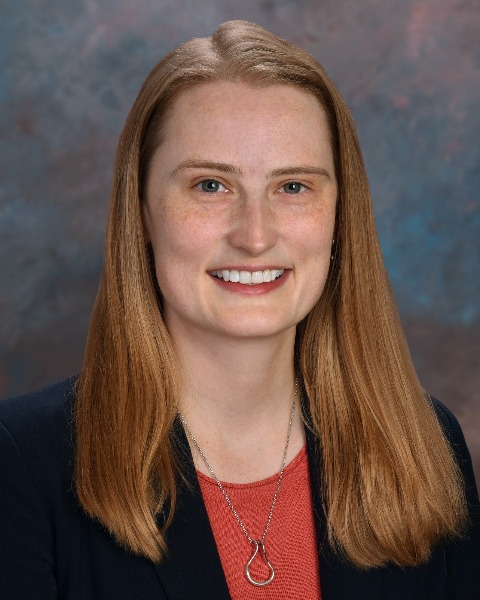
Primary Investigator: Dr. Neha S Dangayach, Assistant Professor of Neurosurgery and Neurology, Icahn School of Medicine at Mount Sinai

Background: Aneurysmal subarachnoid hemorrhage (SAH) is a public health problem affecting 30,000 American adults every year with high mortality and substantial morbidity amongst survivors. Patients often need to undergo inter-facility transfers (IFT) to ensure timely access to resource-intensive, life-saving treatments, which are available only at designated sites.The phases of care for stroke patients may include the following phases: pre-hospital, emergency room (ER), IFT, in hospital, post-acute, and long-term recovery. "Fragmentation" in healthcare delivery means the systemic misalignment of incentives or lack of coordination that adversely impacts care quality, medication errors, resource utilization, cost, and outcomes, and there is no empirically informed guidance on how to integrate these phases as of now.

A learning health system approach allows health systems to leverage the electronic health record (EHR) to implement process improvements and to provide better care for future patients. Such an approach has reduced fragmentation of care in patients with heart failure but has not been studied for stroke patients. The promise of seamless integration of data for patient care via the EHR has not been realized due to several barriers, but the persistence of these barriers despite the availability of tools to overcome them is unacceptable for time sensitive disease processes such as stroke where “Time is Brain”.

Dr. Dangayach’s team will evaluate the usability and adoption of a learning health system and dashboard by different stakeholders. Although the EHR and analytic tools exist, they surprisingly have never been integrated to facilitate informed clinical decisions and implement a learning health system for one of the most fatal health conditions.

Research Objective: The goal of Dr. Dangayach’s proposal is to leverage a learning health system approach to prevent fragmentation of care for SAH patients by  first creating an EHR agnostic platform and then by building an EHR based dashboard to better inform patients, families and care providers about key milestones and areas of care that can be improved upon in real-time.

Outcomes: Using this grant from the Aneurysm and AVM Foundation, Dr. Dangayach’s team hopes that their research will be the first step in seamless integration of care for SAH patients that will provide new insights, incorporate smart analytics, machine learning to provide better care, clinical decision support and improve short-term and long-term outcomes for SAH patients and improve ways of communication with patients and families via dedicated dashboards.

Source: Dr. Neha S Dangayach, Assistant Professor of Neurosurgery and Neurology, Icahn School of Medicine at Mount Sinai. This research summary has been adapted and edited from Dr. Dangayach’s research proposal.


Research Study: Implementing Auricular Trancutaneous Vagal Nerve Stimulation Following Subarachnoid Hemorrhage To Modulate Neuroinflammation And Improve Outcome
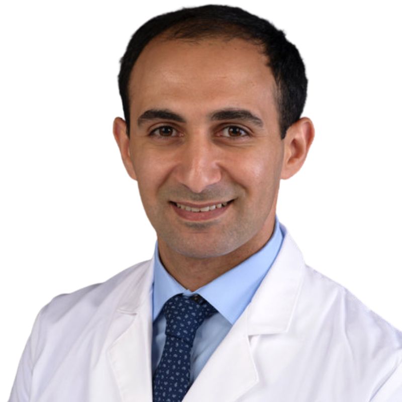
Primary Investigator: Dr. Anna Huguenard, Washington University in St. Louis, Department of Neurosurgery

Background: Intracranial aneurysms are common, with 3 to 5% of adults having at least one. Presence of an intracranial aneurysm is associated with a risk for rupture, which leads to a type of bleed called a subarachnoid hemorrhage (SAH). In those people who develop a SAH, there is a high risk of death or permanent disability as a result. In addition to the immediate effects from SAH, there are other  associated complications which include buildup of fluid in the brain (hydrocephalus), early brain injury and swelling, and reduced blood flow through the vessels in the head (cerebral vasospasm), that all contribute to poor outcomes in these patients.

Aneurysm formation and rupture is complex, and highly influenced by genetic and environmental factors. Inflammation throughout the body and locally may lead to aneurysm formation and rupture, as well as worse outcomes following SAH. Several studies have shown there are increases in inflammatory markers within the blood, cerebrospinal fluid, and the brain tissue itself, with evidence that inflammatory markers are correlated with patient outcomes. Other clinical trials have attempted to use medications that target inflammation to treat these patients, but these medications often had side effects or failed to lead to improved outcomes, and therefore have not been used more broadly.

Vagal nerve stimulation (VNS) has known anti-inflammatory effects, and has been successfully demonstrated in other models of  inflammatory conditions like rheumatoid arthritis, sepsis, and inflammatory bowel diseases. Harnessing its anti-inflammatory effects, VNS has also been used in a mouse model of cerebral aneurysms and SAH. In this study, VNS reduced the rupture rate of intracranial aneurysms, the severity of hemorrhage if rupture occurred, and improved survival and outcome after SAH. Given the promising results in animal studies of SAH, Dr. Huguenard proposes to study VNS in her hospital’s SAH population.

Research Objective: The goal of Dr. Huguenard’s proposal is to study VNS in their hospital’s SAH population, as well as studying patients’ outcomes and complications during their hospital stay, and during their recovery after discharge. Samples of blood and cerebrospinal fluid will be  collected throughout a patient’s hospital stay to evaluate changes in inflammatory markers.

Outcomes: With this grant from the Aneurysm and AVM Foundation, Dr. Huguenard’s team hopes that this new approach could provide a safe, affordable, and effective way to reduce the inflammatory response to SAH, and improve patient survival and recovery. Despite the high risk for death or injury from SAH, there are few consistent treatments to improve outcomes in these patients, so this research could be a huge benefit to recovering patients.

Source: Dr. Anna Huguenard, Washington University in St. Louis, Department of Neurosurgery. This research summary has been adapted and edited from Dr. Huguenard’s research proposal.


Research Study: Assessment of RNA Expression of Adhesion Molecules, Mechanoreceptors, and Inflammatory Proteins in Endothelial Cells Harvested From Unruptured Intracranial Aneurysms in Adults.

Primary Investigator: Zaid Aljuboori, MD, Department of Neurosurgery, University of Washington

Background: Intracranial aneurysms are acquired lesions with a genetic predisposition in certain patient groups. They may present with subarachnoid hemorrhage which is the most feared complication as it commonly results in multiple clinical sequala such as stroke, cognitive dysfunction, and others and leaves 50% of patients permanently disabled. Intracranial aneurysms have a  prevalence rate of about ~2% and are commonly detected on brain imaging performed for unrelated reasons. The exact pathophysiology for aneurysm development, growth, and rupture remains elusive. Existing evidence suggests that aneurysm formation and growth result from a complex interaction among several factors including patients’ genetic profile, vascular anatomy, hemodynamic characteristics, and environmental factors.

Endothelial cell surface mechanosensitive molecules (MSM) (e.g., integrins, caveolar signalosomes, and mechanosensitive ion  channels) are responsible for mechanotransduction and cell adhesion. Several animal studies have alluded to the role of mechanosensitive molecules in the development of vascular malformations such as intracranial or aortic aneurysms. To date, there are no studies that have systematically evaluated the RNA expression profiles of a wide array of mechanosensitive molecules in patients with unruptured intracranial aneurysms. Assessment of the changes of these molecules in unruptured intracranial aneurysms through studying their RNA expression is crucial to understanding their role in the development and evolution of intracranial aneurysms and may lead to the inception of new diagnostic and or therapeutic modalities.

PAI-1 is a protein that plays an important role in fibrinolysis and extracellular proteolysis. It inhibits plasminogen activation by tissue-type plasminogen activator (tPA). TGF, which controls PAI-1 expression, is upregulated in endothelial cells of unruptured cerebral aneurysms. To date, there are no biological markers that can be  easily applied in clinical settings to quantify the degree of inflammation within an aneurysm wall. Since PAI-1 is upregulated in inflammation, it’s conceivable that endothelial cells of unruptured intracranial aneurysms will show an increased expression of PAI-1. Moreover, PAI-1 levels can be measured in plasma and can potentially be used as a biomarker to quantify the degree of inflammation within the aneurysm wall and to assess the effects of therapeutic interventions aimed at alleviating aneurysm wall inflammation.

Research Objective: The goal of Dr. Aljuboori’s proposal is to compare the differences of RNA expression profiles of PAI-1 and adhesion molecules/mechanoreceptors (Primary cilium, Polycystin 1, E- & P- selectins, TRPM7, and TRPV4) between endothelial cells harvested from unruptured intracranial saccular aneurysms and  peripheral arteries (e.g., radial or femoral). 

Outcomes: This grant from the Aneurysm and AVM Foundation will allow for the completion of this pilot study and the obtained data will be used to support a larger grant application to study the molecular pathways that govern the expression of the target molecules in aneurysmal vascular endothelial cells to explain their role in aneurysm development and evolution.

Source: Zaid Aljuboori, MD, Department of Neurosurgery, University of Washington. This research summary has been adapted and edited from Dr. Aljuboori’s research proposal.

The Aneurysm and AVM Foundation is pleased to announce the recipients of the 2020 Cerebrovascular Research Grant Awards. We selected two researchers whose scientific projects showed the greatest potential to improve our understanding of cerebrovascular diseases.

Research Study: Evaluating the use of Remote Ischemic Preconditioning in Patients Undergo Endovascular Repair of Brain Aneurysms
Primary Investigator: John W. Thompson, Assistant Scientist, Neurological Surgery, UMCVI Director of Basic Science Research, University of Miami, Miller School of Medicine

Background: Cerebral aneurysms are common with a prevalence of 2-3% in the general population and exceeding 19% in high-risk populations. Cerebral aneurysm rupture produces a devastating form of stroke resulting in high rates of mortality and morbidity. Endovascular treatment of cerebral aneurysms is becoming the first-line treatment modality for both ruptured and unruptured CA. However, procedures in which catheters are introduced into the cervical carotid arteries commonly dislodge embolic materials that may lead to ischemic damage of varying frequency and severity.

A majority of patients undergoing endovascular aneurysm repair suffer from procedurally-induced infarcts, which are typically small-volume lesions, however some of these infarcts are immediately clinically evident as motor or sensory deficits or neurocognitive impairment. Other infarcts are clinically silent in the short-term but may manifest in long-term consequences such as early onset vascular dementia, and Alzheimer’s Disease. There are currently very limited treatments to reduce the risk of embolic infarcts during endovascular procedures.

Dr. Thompson believes remote ischemic preconditioning (RIPC) might be a key strategy in reducing the number of procedurally induced embolic infarcts. RIPC has been used for protection against both ischemic and reperfusion injury throughout the body, however it has never been used for protection against both ischemic and reperfusion injury throughout the body. Given the high frequency of embolic infarcts occurring during aneurysm repair and the potential for these to induce neurological alterations, this proposal will test this novel therapy to directly address this unmet need in patients undergoing endovascular aneurysm repair.

Research Objective: The goal of Dr. Thompson’s research is to define the molecular events behind cerebral aneurysm formation, growth and rupture and to develop novel medical therapies for the successful treatment of cerebral aneurysm patients. Currently, there are very limited medical therapies to prevent or reduce this issue in endovascular treated patients, so this study will investigate the potential of remote ischemic preconditioning as a therapeutic intervention for endovascular procedure induced embolic ischemic events.

Outcomes: With this grant from The Aneurysm and AVM Foundation, Dr. Thompson and his team will conduct their research to contribute to a better understanding of the efficacy of RIPC in the prevention of endovascular induced embolic infarcts as well as the mechanisms through which RIPC may lead to neuroprotection. He hopes that the positive impact of this research can enhance our understanding of RIPC neuroprotection and will give clinicians a new medical therapy to better patient outcomes following endovascular treatment of cerebral aneurysms.

Source: John W. Thompson, Assistant Scientist, Neurological Surgery, UMCVI Director of Basic Science Research. This research summary has been adapted and edited from Dr. Thompson’s research proposal.


Research Study: Neutrophil Infiltration in Coiled Aneurysm Healing: Adjuvant IL-17 Blockade

Primary Investigator: Koji Hosaka, Associate Scientist, University of Florida Division of Sponsored Programs

Background: Cerebral aneurysms affect up to 5% of the population. When aneurysms rupture, they cause devastating subarachnoid hemorrhage (SAH), which accounts for 5% of strokes and has an estimated mortality of 50%. Current treatment options to prevent rupture or rebleeding include surgery (craniotomy and clipping) or endovascular treatment (coiling or flow diversion stenting). Endovascular coiling appears to be safer than surgery for unruptured and ruptured aneurysms, however, coiling is associated with up to 44% incomplete treatment.

Based on surgical/autopsy studies, durable cerebral aneurysm healing occurs when a durable tissue plug containing immune cells, collagen, smooth muscle cells, and angiogenesis persists  in the aneurysm sac. As thrombus dissolves, aneurysm recurrence may occur unless fibrotic tissue has already filled the spaces between the implanted coils. Polymer coated coils, although intended to increase aneurysm filling density, have not shown efficacy in clinical trials, suggesting a fundamental gap in knowledge regarding the mediators and mechanisms of action of durable aneurysm healing.

Dr. Hosaka’s lab has shown that osteopontin (OPN) sustain-releasing coils produce a robust tissue ingrowth response, and has found that IL-17(A) is transiently expressed in OPN-coiled aneurysms at  day 1 following coil treatment. The proposed anti-IL-17 therapy would intend to mitigate aneurysm recurrence with coiling. They will further investigate the relationship between duration of IL-17 blockade and neutrophil recruitment in the context of OPN-mediated murine aneurysm healing.

Research Objective: The goal of Dr. Hosaka’s proposal is to assess feasibility and potential duration of anti-IL-17 neutralizing antibody as an ancillary treatment in the context of aneurysm coiling, and to gain insight into the aneurysm healing cascade. This will be achieved through evaluating the temporal cascade of IL-17 blockade on neutrophil infiltration in OPN-coiled aneurysm healing, as well as determining the optimal duration of IL-17 blockade treatment for durable aneurysm healing. 

Outcomes: Using this grant from the Aneurysm and AVM Foundation, Dr. Hosaka and his team will work to facilitate the long-term goal of their research to improve treatment for cerebral aneurysms by defining the process of aneurysm healing and employing agents to accelerate and improve that process. This work is directly translatable and deliverable to patients, and future directions after establishing basic mechanisms of aneurysm healing in the mouse model will be to test in a rabbit aneurysm model, the direct precursor to human trials. 

Source: Koji Hosaka, Associate Scientist, University of Florida Division of Sponsored Programs. This research summary has been adapted and edited from Dr. Hosaka's research proposal.

The Aneurysm and AVM Foundation is pleased to announce the recipients of the 2019 Cerebrovascular Research Grant Awards. We selected two researchers whose scientific projects showed the greatest potential to improve our understanding of cerebrovascular diseases.

Research Study:  Human Genetics and Molecular Mechanisms of Vein of Galen Malformation

Primary Investigator:  Kristopher Kahle, MD, PhD., Assistant Professor of Neurosurgery, Pediatrics, and Cellular & Molecular Physiology, Yale School of Medicine; Yale-Rockefeller NIH Center for Mendelian Genomics; Director, Developmental Anomaly Neurosurgery, Yale-New Haven Hospital

Background: Vein of Galen malformation (VOGM) is one of the most common and devastating forms of arteriovenous malformations (AVMs) in children. Despite advances in treatment in recent years, VOGM still frequently leads to heart failure, cerebral fluid imbalance, bleeding within the brain, and even death among affected children. Beginning in infancy, many children undergo multiple surgeries, scans, and invasive procedures, all of which cause enormous physical, emotional, and financial strain on patients and their families.

Very little is known about the causes of VOGM. This lack of knowledge has hindered the development of new and improved methods to diagnose and treat this disease. These obstacles can be overcome using whole exome sequencing (WES), an approach that has led to unprecedented gene discoveries in other structural brain disorders.

He hypothesizes that mutations in interacting components of an EPHB4 signaling network contribute to VOGM by disrupting RASA1/mTORC1-dependent arteriovenous specification. Furthermore, Dr. Kahle hypothesizes that phenotypic heterogeneity and incomplete penetrance of VOGM and variable expressivity may be explained by the compound effect of inherited germline mutation and with tissue-specific somatic mutations.

Research Objective: The goal of Dr. Kahle’s research is to fill these knowledge gaps using the latest in DNA-sequencing technology. Utilizing the power of social media, Dr. Kahle has connected with and recruited patients from around the globe to participate in his genetic study creating the largest cohort of VOGM patients to date. He expects to expand that VOGM case-parent cohort, more than doubling his initial study. He also expects to screen for known VOGM-associated mutations as well as carry out whole exome sequencing and bio-informatic analysis. Finally, Dr. Kahle expects to analyze somatic vascular tissue from germline-mutant VOGM patients for superimposed somatic mutations.

Outcomes: With funds from The Aneurysm and AVM Foundation, Dr. Kahle believes that his approaches will expand the current understanding of how VOGM develops at the molecular level. He hopes that the resulting advances will improve disease management, surveillance, and genetic counseling, stimulate research into new diagnostic and therapeutic options, and improve social support for patients and families afflicted with VOGM and possibly other brain and systemic AVMs.

Included in Dr. Kahle's award is the Lauren Karafiol Cerebrovascular Research Grant. As part of TAAF's part of annual grant program, this award is made in memory of Lauren P. Karafiol and aims to off-set costs of research in the area of AVMs, the disease that forever changed Lauren's life and that of her family. We are incredibly proud to join Lauren's family in support of novel research of arteriovenous malformations.

Source: Kristopher Kahle, MD, PhD, Yale University School of Medicine. This research summary has been adapted and edited from Dr. Kahle’s research proposal.


Research Study:  Patient-specific Genetics of Cerebral Aneurysm Endothelium
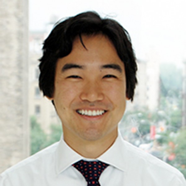
Primary Investigator:  Michael R. Levitt, MD, Associate Professor of Neurological Surgery and Radiology, University of Washington. Dr. Levitt holds several other titles with UW, chiefly Scientific Director of the Stroke and Applied Neuroscience Center.

Background: Cerebral aneurysm formation, growth and rupture occur as a result of forces of blood flow on the endothelial cells that line the arteries in the brain. These forces, called “hemodynamic stresses,” can be calculated from computer simulations, but their effects on endothelial cells cannot be determined in actual patients. In fact, a major drawback of existing CFD (computational fluid dynamics) studies in clinical applications (such as risk of aneurysm growth or rupture, or outcome of aneurysm treatment) is that they do not directly measure the pathological effects of hemodynamic variables on biological tissue. Rather they inferred from a variety of sources such as animal studies and in vitro experiments. The current state of the art cannot provide clinicians with the ability to measure the cellular response of the cerebrovascular endothelium to hemodynamic stress in a specific patient’s cerebral aneurysm, which is a significant gap in knowledge.

To address this critical knowledge gap, and to better characterize the effect of hemodynamic stress on aneurysm endothelial cells, Dr. Levitt’s research will use 3D-printed models of patient’s cerebral aneurysms, which are coated with living human endothelial cells and exposed to hemodynamic stress. The genetic and protein response of these cells to different amounts of hemodynamic stress will be measured.

Research Objective: The goal of Dr. Levitt’s research is to further our understanding of the patient-specific hemodynamic, genetic, and proteomic factors that contribute to aneurysm formation, growth, rupture, and healing thus informing treatment decision-making and improving the quality of life for patients suffering from intracranial aneurysms.

Outcomes: With funds from The Aneurysm and AVM Foundation, Dr. Levitt believes that his translational approach bridges the gap between computational fluid dynamics and vascular biology and intends to produce a quantified reference database of endothelial expression of key vascular factors in response to differential levels of hemodynamic stress. The output of this proposal can then be applied to future CFD simulations of aneurysms and permit an informed estimate of the endothelial response to a given level of hemodynamic stress. Dr. Levitt believes these findings will link computer simulations with actual endothelial cell activity, which will allow clinicians to better predict aneurysm risk, and develop new treatments such as gene therapy and targeted drugs to treat cerebral aneurysms without surgery.

Source: Michel R. Levitt, MD, University of Washington. This research summary has been adapted and edited from Dr. Levitt’s research proposal.

Research Study:  Mechanisms of Neurocognitive Decline After Subarachnoid Hemorrhage

Primary Investigator:  David Y. Chung, M.D., Ph.D., Instructor in Neurology at Harvard Medical School, Division of Neurocritical Care and Emergency Neurology at Massachusetts General Hospital

Background: Subarachnoid hemorrhage (SAH) from a ruptured brain aneurysm is a life-altering condition that affects more than 30,000 Americans and costs $5.6 billion annually. Approximately 20% of patients who survive the initial rupture will go on to have subsequent, secondary brain injury. Furthermore, even patients with relatively good outcomes frequently suffer from persistent cognitive deficits precluding return to work. Both the pathophysiology underlying secondary brain injury and the mechanisms underlying lasting cognitive deficits remain unknown.

Emerging human evidence suggests that cognitive deficits following SAH are associated with altered functional brain connectivity. Moreover, there is strong human evidence that spreading depolarizations (SD)—similar to the phenomenon of spreading depression in migraine aura—are associated with secondary injury and cognitive decline after
SAH7-13. SDs are thought to arise out of ischemia14, but it is difficult to distinguish the individual contributions of SDs and ischemia to outcome using existing animal models. Therefore, we will use novel mouse models of SAH to examine the contribution of SDs in the absence of ischemia on functional brain connectivity and neurocognitive decline.

Research Objective: The goal of Dr. Chung's research is to prevent further brain damage after aneurysm rupture and to develop therapies that help survivors recover back to their baseline function. In particular, he is trying to understand why some patients who look well after surgical repair of an aneurysm go on to develop a poorly understood syndrome of progressive brain injury and functional decline. At the same time, he is trying to understand why others who do well after their hospitalization have long-lasting cognitive problems that prevent return to work.

Dr. Chung believes that a phenomenon called spreading depolarization causes further injury after aneurysm rupture by changing how different parts of the brain connect to each other. To study spreading depolarizations and connectivity, he has developed a set of non-invasive tools to experimentally cause spreading depolarizations in an animal model of aneurysm rupture. Furthermore, he has developed ways to determine how difference parts of the brain connect to each other using a cutting edge technique in living mice called resting state optical intrinsic signal imaging.

Outcomes: With funds from The Aneurysm and AVM Foundation, Dr. Chung believes that the findings from his research will be important for understanding why survivors of aneurysm rupture continue to develop brain injury in the hospital and suffer persistent long term cognitive deficits. The greatest hope is that knowledge gained from his work will lead to a breakthrough in the field and directly lead to new therapies for people who have had a ruptured brain aneurysm.

Source: David Y. Chung, M.D., Ph.D., Massachusetts General Hospital and Harvard Medical School . This research summary has been adapted and edited from Dr. Chung's research proposal.

Research Study:  Histological and Blood Flow Evaluations of AVM and Cerebral Artery Vasculature to Create a Simple Computational Fluid Dynamic Model of Arteriovenous Malformations
Primary Investigator:  Nina Moore, MD, MSE, Dept. of Neurosurgery at the Cleveland Clinic Foundation

Background: Carrying a 3% risk of hemorrhage per year, cerebral arterial venous malformations (AVMs) pose a difficult question to physicians who need to decide whether to treat the AVM or monitor conservatively as was recently suggested in the ARUBA trial.  With a paucity of prospective studies that stratify the risk of AVM rupture based on specific anatomic features, physicians have to piece together outcome studies that may not fir their patient’s AVM.  It would be clinically useful to have the ability to accurately predict whether a patient’s particular AVM anatomy would predispose them to rupture and the timeframe in which to expect rupture.

Computational fluid dynamics (CFD) is a promising technique for modeling the human vascular system and examining vascular disease processes.  Models of the cardiac anatomy and cerebral aneurysms with CFD are adding insight into the hemodynamic changes the vessels undergo. CFD models can illuminate risk factors with particular shape, sizes, and flow patterns seen in aneurysms and vascular malformations as different stresses affect the vessels.  This knowledge can significantly expand when CFD is coupled with structural analysis of blood vessel walls providing a more comprehensive way to evaluate cerebrovascular disease.  To date, this technique has not been applied to modeling of cerebral AVMs.

Research Objective: Utilizing the field’s current understanding of computational fluid dynamics applied to cerebral aneurysm and blood vessels, the object of this research is to develop a simple model of an AVM using properties defined by histologic analysis of cerebral blood vessel wall structure as well as resected arteriovenous malformation vessels.  Additionally, the work will seek to obtain intraoperative and angiographic comparisons of velocity within the arteriovenous malformations to correctly simulate arteriovenous malformation flow physiology.  Specifically, Dr. Moore hypothesizes that they can create a simple AVM model within a computational fluid dynamics program that incorporates accurate anatomical wall structure properties and accurate flow parameters.  This model could then be later evaluated to predict the parameters of distension and failure of the vascular malformation wall.  The long term goal of this project is to develop a personalized medical approach to a patient’s unique AVM.  The hope is that the information learned from these simulations would serve as the groundwork for the development of a tool that allows for testing of different treatment strategies—embolization, surgery, radiosurgery or conservative therapy, eventually allowing the surgeon to even test details of their approach for treatment of an AVM.

Outcomes: With funding from The Aneurysm and AVM Foundation, Dr. Moore and her team will work in three phases.  The first phase will be cerebrovascular wall histological studies, followed by phase two consisting of the study of blood flow rate in live AVMs, and finally phase three which will be the creation of the computational fluid dynamics model.  Utilizing the date from this study, Dr. Moore hopes to progress towards building a mathematical model that accurately predicts the natural history of rupture in AVMs giving surgeons and patients a roadmap to better treatment strategies.

Source: Nina Moore, MD, MSE the Cleveland Clinic Foundation. This research summary has been adapted and edited from Dr. Moore's research proposal.


Research Study:  Ceruloplasmin concentration and ferroxidase activity in CSF and risk of brain injury after aSAH
Primary Investigator:  Joao Gomes, MD (PI), Assistant Professor of Medicine (Neurology), Neurointensivist and Director of the neuro-ICU at Cleveland Clinic; Leah P. Shriver, PhD (Co-I), Assistant Professor at the University of Akron in the Department of Chemistry; and Christopher J. Ziegler, PhD (Co-I, Professor at the University of Akron in the Department of Chemistry

Background: There is accumulating evidence that iron-mediated brain injury and oxidative damage contribute to poor outcomes following aneurysmal subarachnoid hemorrhage (aSAH).  Because of its ferroxidase action, the protein ceruloplasmin (Cp) regulates iron levels in the central nervous system and prevents free radical injury. 

Paradoxically, reactive oxygen species can bring about modifications in the structure of Cp that result in decreased ferroxidase activity, potentiating a vicious cycle of oxidative stress.

Research Objective: The objective of Drs. Ziegler, Shriver, and Gomes’ research is to examine the relationship between Cp concentration and its ferroxidase activity in cerebrospinal fluid and the development of delayed cerebral ischemia and neuronal cell injury following aSAH.  Furthermore, they want to determine if CNS Cp undergoes oxidation and structural modifications following aSAH that result in decreased enzymatic activity.

This represents a novel pathway for aSAH pathogenesis and a promising potential therapeutic target that thus far remains unexplored.

Outcomes: With funding from The Aneurysm and AVM Foundation, Drs. Ziegler, Shriver and Gomes hope to showcase that Cp has a protective effect following aSAH and that its concentration and ferroxidase activity in CSF are inversely associated with the development of DCI. Furthermore, we hypothesize that the oxidative milieu present in the CSF of high grade aSAH patients will lead to modifications in the protein structure of the Cp molecule that will in turn result in decreased enzymatic activity and higher risk of DCI.

Source: Joao Gomes, MD the Cleveland Clinic. This research summary has been adapted and edited from Dr. Gomes’ research proposal.

Research Study:  An Investigation of Epoxyeicosatrienoic Acids as a Treatment Strategy to Improve Glymphatic Flow, Cerebral Blood Flow and Behavioral Outcomes Following Subarachnoid Hemorrhage in Rats
Primary Investigator:  Tristan Stani, MD, Dept. of Neurological Surgery at Oregon Health and Science University

Background: Aneurysmal subarachnoid hemorrhage (SAH) remains one of the most challenging stroke syndromes facing today's neurological critical care providers and surgeons.  Despite advances in characterizing the anatomical details of aneurysms and the continued development of novel surgical and endovascular techniques for securing aneurysms, the delayed consequences of initial aneurysm rupture continues to challenge care providers,  We continue to lack a sophisticated understanding of the exact systems that are disrupted following SAH and the mechanisms that underlie their dysfunction.  All too often the most technically successful treatment of a ruptured aneurysm is unfortunately marred by the delayed stroke and other cerebral dysfunction for which we have limited treatment options

Research Objective: The central objectives of this project are to determine the effectiveness of augmented levels of epoxyeicosatrienoic acids on rescuing perivascular flow of CSF through the glymphatic system in the setting of subarachnoid hemorrhage and to correlate post-SAH changes in the glymphatic system and subsequent glymphatic flow rescue by epoxyeicosatrienoic acids with post-SAH cerebral blood flow changes and behavioral outcomes. 

The “glymphatic system” is a cerebral microcirculation system of cerebral spinal fluid (CSF) that has only recently been described. Initial work on this system has suggested important connections with neurodegenerative diseases such as Alzheimer’s Disease. Importantly, the system has also recently been demonstrated to be disrupted following SAH and then restored after the delivery of tPA, a “clot-busting” drug commonly given in the clinical setting following acute ischemic stroke. This suggests important new treatment strategies for SAH.

Outcomes: With funding from The Aneurysm and AVM Foundation, Dr. Stani will launch a series of MRI imaging experiments which will characterize glymphatic CSF flow pre- and post-SAH.  He will then image glymphatic flow pre and post-SAH in an experimental group pretreated with analogs to epoxyeicosatrienoic acids (EETs).  EETs are endogenous molecules that the body already makes which possess profound anti-inflammatory, vasodilatory and fibrinolytic effects.  He hopes to demonstrate that the fibrinolytic  ("clot-busting") effects of EETs analogs will improve glymphatic CSF microcirculation and ultimately improve cerebral blood oxygen delivery and decrease cerebral inflammation following SAH, which will ultimately lead to improved outcomes.

Source: Tristan Stani, MD, Oregon Health and Science University. This research summary has been adapted and edited from Dr. Stani's research proposal.


Research Study:  Quality of Life in Patients Diagnosed with Unruptured Cerebral Aneurysm: Prospective Single-Center Series

Co-Primary Investigators:  Peter Gooderham, MD, Clinical Assistant Professor, Division of Neurosurgery, Dept. of Surgery at the University of British Columbia and Charlotte Dandurand, MD, Neurosurgery Resident at the University of British Columbia

Background: In the United States, 6 million people, 2% of the population, are living with an unruptured brain aneurysm.  Many of these people are unaware of their diagnosis.  The most important and devastating consequence of a brain aneurysm is subarachnoid hemorrhage from aneurysm rupture. Living with the diagnosis of an unruptured cerebral aneurysm can understandably cause anxiety for a patient and the impact on patients' quality of life is not well understood. The degree to which this diagnosis affects patients and how this affect changes over time remains unknown.  The impact of microsurgical clipping and endovascular coiling on patients' quality of life is also poorly studied

Research Objective: The objective of this research is to identify how the diagnosis of an unruptured cerebral aneurysm and its subsequent treatment impacts quality of life over time.

Outcomes: With funding from The Aneurysm and AVM Foundation, Dr.'s Gooderham and Dandurand will use objective quality of life tools to interview patients at diagnosis and again one year later.  Quality of life will be assessed at diagnosis, at 6-12 weeks post-operative follow-up, and at 1 year post-operative follow-up in patients who have been treated.  The latter group will be divided into 2 sub-groups, endovascular and microsurgical (clipping). Patient demographics, size and location of aneurysm, radiological features, presence and degree of neurological deficits, treatment modalities, and postoperative complications will also be collected.

Source: Charlotte Dandurand, MD, University of British Columbia. This research summary has been adapted and edited from Dr.'s Gooderham and Dandurand's research proposal.